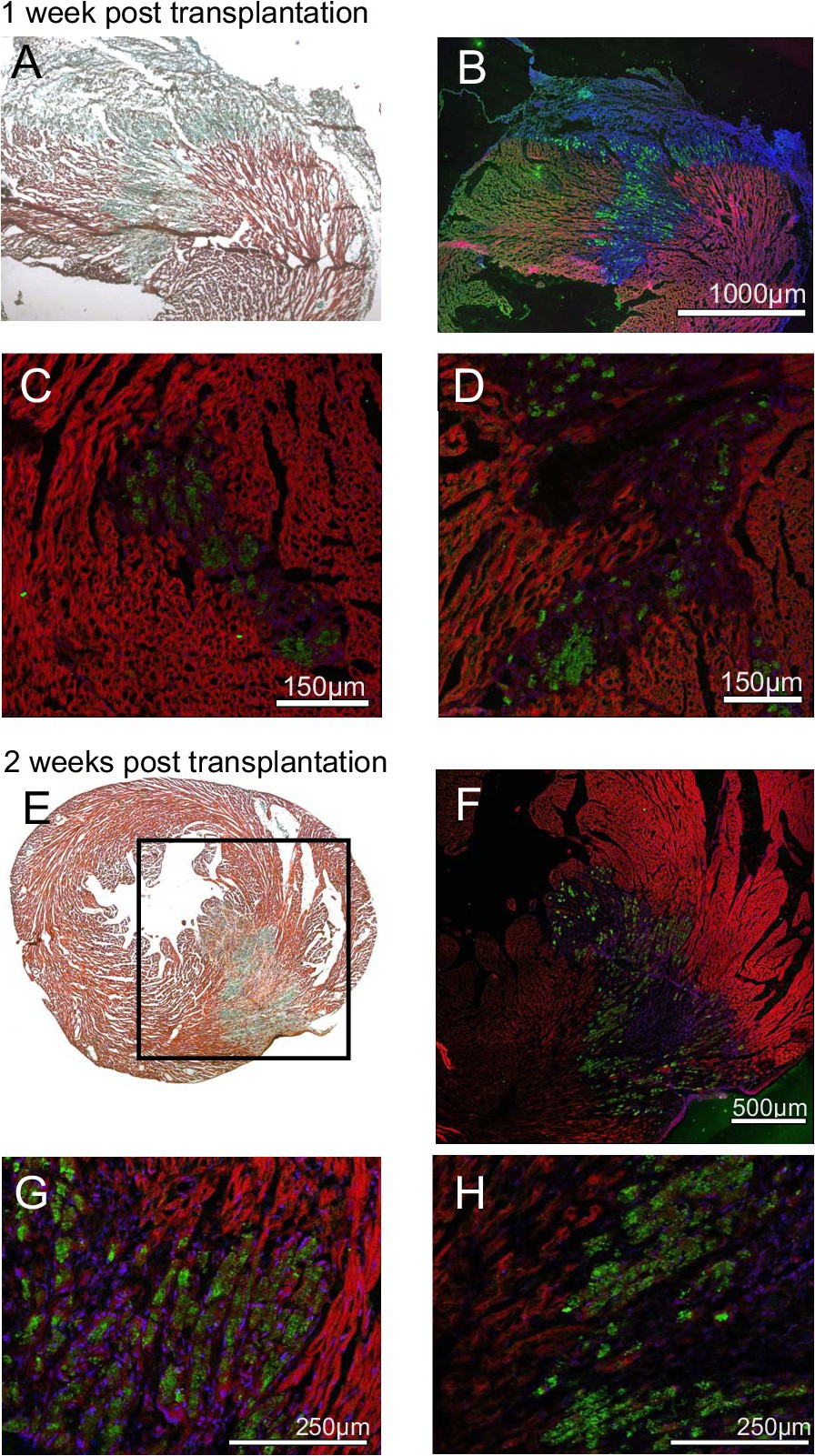
Fig. 5. Histological observation of transplanted micro-tissues. Micro-tissues of low-dose MSC/iPS-CM were transplanted into cryoinjured cardiac tissue. Trichrome staining revealed fibrotic areas of myocardial damage after one week (A) and two weeks (E). Transplanted cells were stained with antibodies against luciferase (green) and nuclei were co-stained by Hoechst dye (blue). Tissue autofluorescence (red) was recorded to visualize the endogenous cardiac tissue. Panel A/B and C/D show results for two individual animals one week after cryoinjury and cell transplantation. Panel E/F show engrafted cells from one animal two weeks after cell transplantation. Panel G/H display magnified regions from the section shown in F.